Local News
Blue Ridge Wildlife Center Patient of the Week: American Crow
Photos / Blue Ridge Wildlife Center
Your front row seat to a crow’s fracture repair!
Ever wondered how we repair fractures at a wildlife hospital? Scroll though the photos below to learn how this American Crow’s ulna (wing bone) fracture was surgically stabilized.
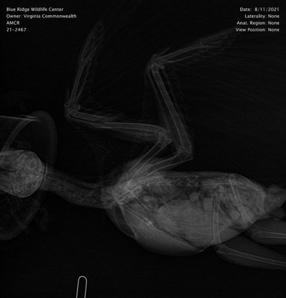
This radiograph shows the American Crow on the day of admission. The left ulna (the thicker bone on the wing that is closest to the head in this image) is fractured. Had the bone been well-aligned, a simple bandage would have been sufficient to stabilize the fracture and allow for healing. In this case, we felt that there would be better healing with surgical stabilization. Wild animals must be able to get around well, find food, and live without chronic pain. The bone simply healing together is rarely good enough. It needs to be fully functional. To give this bird the best chance of long-term survival, surgery was performed.
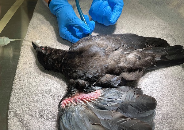
Prior to surgery, the wing was bandaged and the patient was given strong pain medications. Fluids were administered to rehydrate this bird and antibiotics were also started as metal would be placed in this bird’s bones. Once stable for surgery, the patient was placed under general anesthesia using an inhaled anesthetic and intubated so that we could have better control of the airway. In the above photo, the crow is being prepared for surgery and a veterinary student is getting a heart rate.
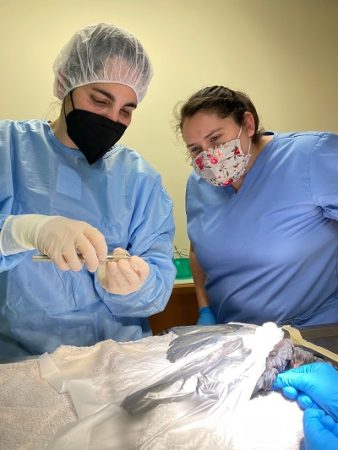
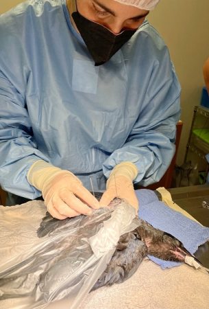
In the first photo above, the crow has been scrubbed and draped (with a clear, sterile, sticky drape that allows us to visualize the bird well throughout surgery). Our veterinarian, Dr. Riley, is explaining how we select surgical pins and how to use the surgical instruments to one of our veterinary externs. In most veterinary schools, very little time is spent on avian medicine and surgery, externships like those provided at Blue Ridge Wildlife Center are an essential first step in the training of avian veterinarians!
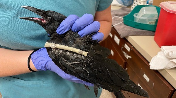
An external fixator was used to align and stabilize the ulna for this crow. This involved placing surgical pins perpendicular to the bone itself, as seen in the second photo.
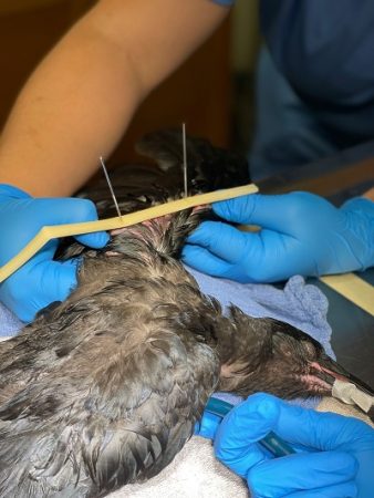
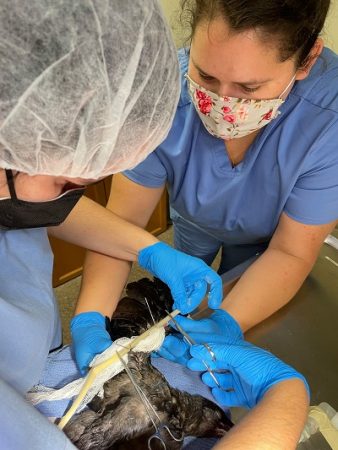
For many animals, an external fixator is completed with a fixator bar – a steel bar that holds the pins in place. Since we have such small patients, we do not need expensive, surgical steel fixator bars. We create our own fixator bars using penrose drains (the cream-colored plastic-like material seen in this photo between pins) and fill the tube with acrylic. This creates a solid bar that acts like a steel bar, holding the bone segments in place while they heal.
In the second photo, one of our veterinary students is holding and keeping the fracture stable while the acrylic material in our “fixator bar” hardens. Once hard, the external fixator will hold those bone segments in place.
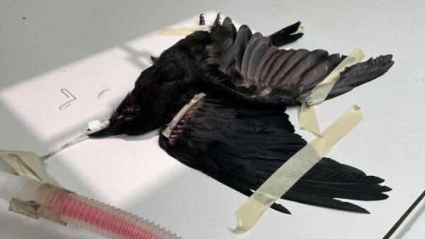
After surgery, follow up radiographs are taken to ensure that the bone segments are positioned well for healing.
One benefit of surgical fixation over bandaging is that the bird can use that limb right away! This prevents the stiff joints and risk of contracture that often come with bandaging. In a bandaged fracture, we sedate patients for regular bandage changes and physical therapy. With external fixators, this is unnecessary as the patient can move and use those joints as soon as they wake up. Active use also helps the bones to heal more quickly!
Mocha
Did you vote for our newest ambassadors name? We are excited to announce that the name ‘Mocha’ is the winner! Come visit Mocha at our Wildlife Walk soon!

4 Stimulating Activities for a Healthy Brain After 50

The First Amendment: America’s Unique Foundation of Freedom

How to Tackle Credit Card Debt

Summer Enrichment Camp Gives Teens Hands-On Learning Opportunities

VDOT: Warren County Traffic Alert for April 6 – 10, 2026
David Silek to Remain in Chairman’s Seat of WC Republican Committee Pending 6th District Appeal Decision

Sales Set to Bloom Like Daffodils This Spring

America 250: The Bookseller Who Helped Save the Revolution

Business Growth Series: Why Good Businesses Still Struggle to Grow

Brownies with Mini Chocolate Easter Eggs

Shenandoah Downs Opens 11th Season April 11 with Tribute to Roger Hammer

We Don’t Know Everything About DNA

How PAAS Came to Dominate the Easter Egg Dye Tradition

Spanberger Signs Bipartisan School-Safety, Student Support Bills Into Law

Melanie J. Pomeroy (1958 – 2026)

David Benjamin Heller (1990 – 2026)
1776 Wasn’t Just About Independence

Three-Tiered System for Urban Agriculture Based on Lot Size Takes Shape at Town Planning Commission Work Session

Child Abuse Awareness Month Brings Focus to Reporting, Prevention in Warren County

National Dental Hygienists Week: Is Your Oral Care Routine Optimal?

The Coin That Traveled Through Time and Space

One “What If?” Question Is Changing the Future of Farming

AA Speaker to Explain Recovery Program at United Methodist Men’s Dinner
Jeannette M. Hyland (1944 – 2026)
